
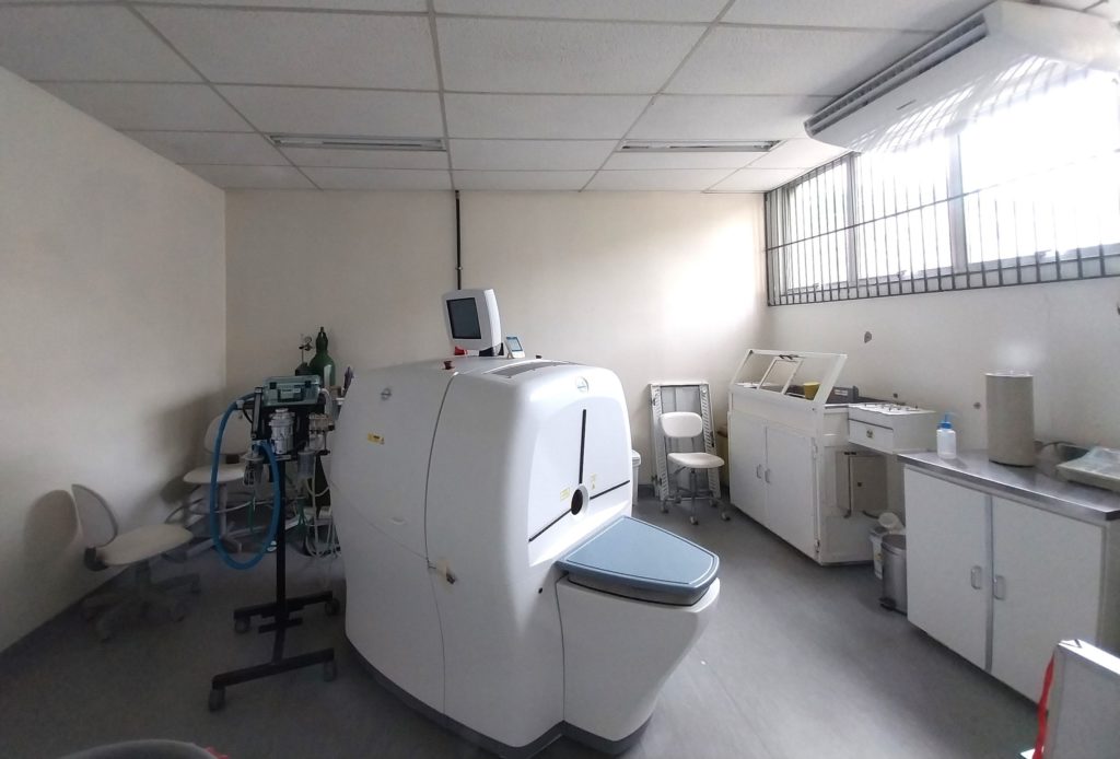
CENABIO-UIPA, located on the ground floor of the UIPA building, has a trimodal equipment (Triumph LabPET 8, Gamma Medica), which integrates three imaging techniques in a single device: PET (positron emission tomography), SPECT (single photon emission tomography) and CT (computed tomography). This combination allows for the rapid and efficient acquisition of anatomical and functional images, facilitating the fusion of information for translational studies..
Computed Tomography (CT)
Imaging technique that uses X-rays to generate 3D images with high spatial resolution, ideal for visualizing bone structures. For soft tissues, the use of contrast agents may be necessary to improve definition.
Molecular Imaging (PET/SPECT)
Functional techniques that detect biological processes at the cellular and molecular level, using radionuclides as metabolic tracers.
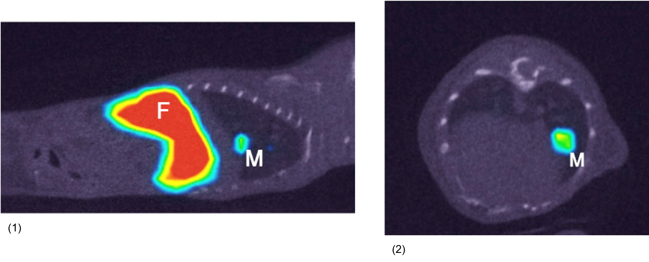
The image is formed by detecting gamma radiation emitted by radiotracers (molecules labeled with radioisotopes), administered in minimum concentrations. Pharmacokinetics and selective uptake by tissues provide quantitative and functional data.
Technical Specifications:
Triumph LabPET8 Gamma Médica (GE)
- CT
- 3D images up to 100 μm.
- PET
- Radionuclide used: 18F;
- Spatial resolution of approximately 1.35 mm.
- SPECT
- Radionuclide used: 99mTc; Spatial resolution of 2 mm to 0.5 mm;
- Two CZT cameras (direct conversion of gamma radiation into electrical signal) and multiple collimators (pinhole, multi-pinhole and parallel).
Image gallery
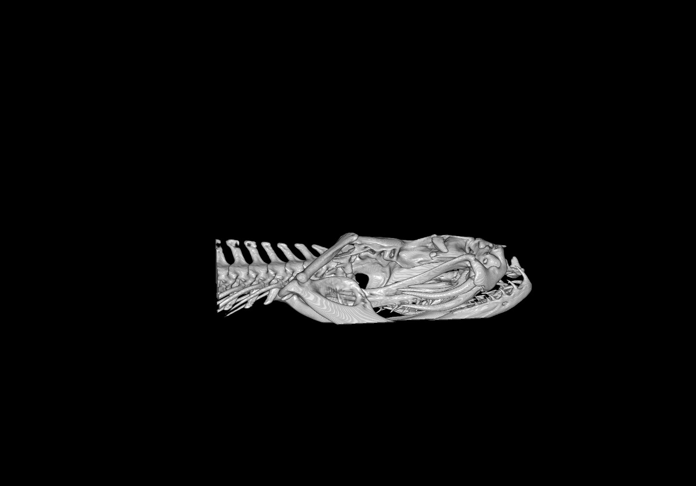
Kindly provided by Breno Hamdan, Instituto Vital Brasil.
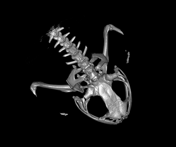
Kindly provided by Fábio Hepp, IB/UFRJ.

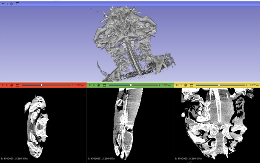
Kindly provided by Karla Soares, IB/UFRJ).
.
Management Committee

Responsible technologist:
CV
Need clarification? Contact us: plataforma.pet.spect.ct@cenabio.ufrj.br


