
FEI QUANTA 250
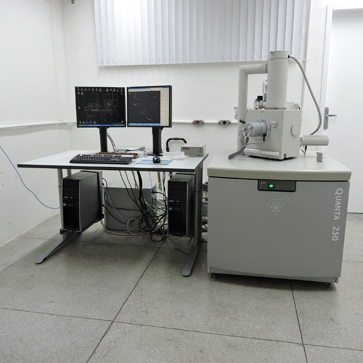
Technical specifications:
Quanta 250 is a scanning electron microscope from FEI (Thermo Fisher). It operates with beam accelerations between 1 and 30 kV and has an Everhart-Thornley secondary electron detector and a backscattering detector. This equipment is able to operate at high vacuum (10-2 to 10-4 Pa), low vacuum (10 to 130 Pa), or environmental vacuum mode (10 to 400 Pa), allowing users to observe even non-conductive and/or hydrated samples (non-coated). Its sample holder has a Peltier Cooling stage for samples sensitive to the electron beam thermal damage. Moreover, it is equipped with a EDAX EDS X-Ray detector (Energy Dispersive Spectroscopy) for micro-analysis and element mapping.
THERMO FISHER QUATTRO S
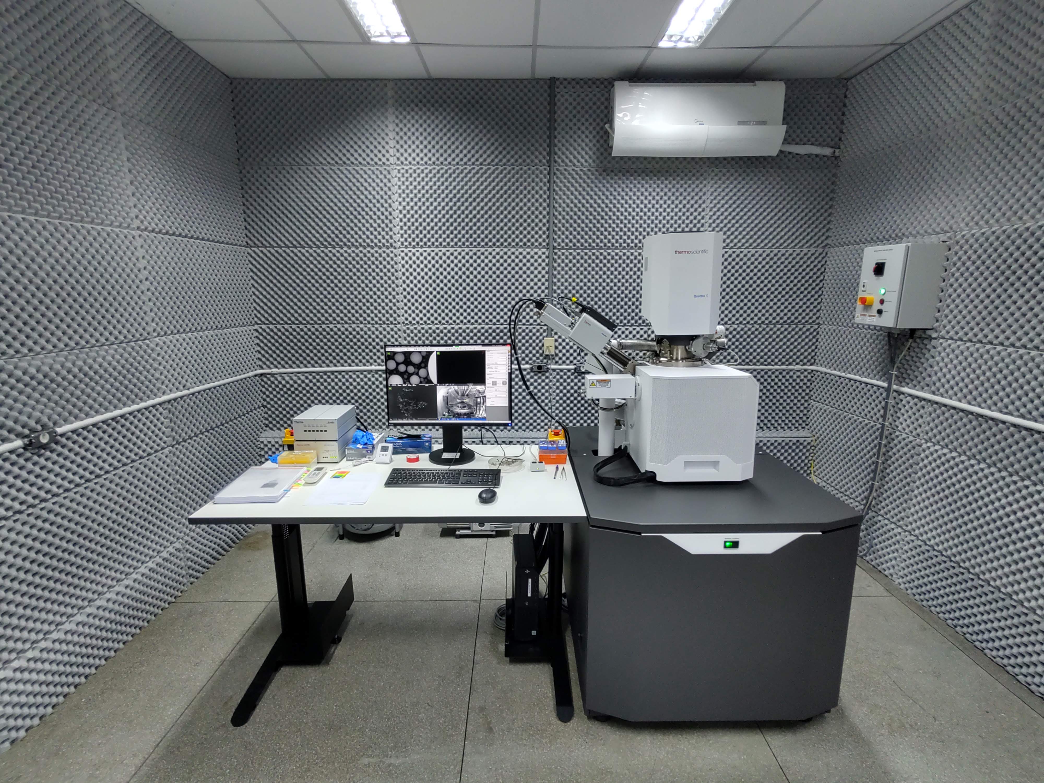
Technical specifications:
Thermo Fisher Scientific scanning electron microscope Quattro S. Operates with accelerating electron voltage between 1 and 30 kV, has secondary electron detectors, backscattered and energy dispersive spectroscopy (EDS) detector. The electron column has a FEG (Field Emission Gun) type source that produces a highly coherent electron beam and with high brightness. It has a beam deceleration system, which enables use at very low voltages (< 1 kV) which allows obtaining high resolution images, at up to 4.0 nm. The equipment can work under high vacuum (10-2 to 10-4 Pa), low vacuum (10 to 200 Pa) or in environmental mode (2600 Pa to 4000 Pa). For observation, samples can be prepared in a traditional way, dried and metallized or observation is allowed on materials in their native state, with non-conductive and/or hydrated materials. The energy dispersive spectroscopy (EDS) detector with element microanalysis identifies the chemical composition of elements quickly generating color maps from scanned images.
ZEISS EVO MA10
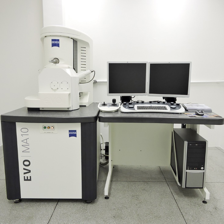
Technical specifications:
EVO MA10 is a scanning electron microscope from Zeiss. It operates with beam accelerations between 1 and 30 kV and has an Everhart-Thornley secondary electron detector and a backscattering detector. This equipment is able to operate at high vacuum (10-2 to 10-5 Pa) or at variable pressure vacuum (10 to 400 Pa), known as VP mode, allowing users to observe even non-conductive (non-coated) samples. On VP mode, the secondary electron detector is a Large Field Detector (LFD).
ZEISS FIB-SEM AURIGA 40
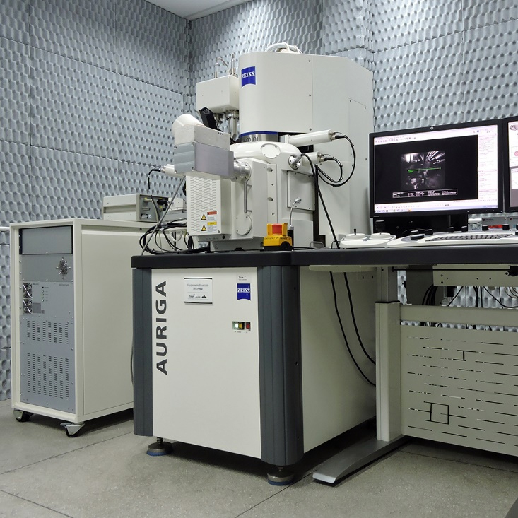
Technical specifications:
High resolution scanning electron microscope and crossbeam (FIB-SEM) built with two columns: an electron beam column (GEMINI) and a focused ion beam column (COBRA); a gas injection system (GIS); and two nano-manipulators. The electron beam column has a field emission gun electron source (FEG) that produces a highly coherent and bright electron beam allowing the user to image at high resolution (» 3.0 nm) and very low-voltage (0.5 to 2.0 kV). The focused ion beam column has a gallium source that produces the focused ion beam, which is primarily used to cut (etching) the sample with great precision. The gas injection system has 5 channels for specific and in-situ tests, four of which are of N2, C, XeF2, and Pt. The 2 nanomanipulators (Kleindiek) have 5 degrees of freedom, which facilitates the handling of the prepared lamella. The microscope is equipped with 2 secondary electron detectors: one for conventional imaging (Everhart-Thornley) and one for ultra-high resolution imaging (In-lens); 2 detectors for backscattered electrons: one retractable solid state detector (NTS BSD) and one for low energy backscattered electrons (ESB); and 1 detector for visualization in STEM mode (Scanning Transmission Electron Microscopy). The dual-beam system (FIB-SEM) allows the user to do Volume EM by using the FIB to cut the sample and the SEM to image in a series of slice-and-view sequence. Additionally, the microscope has the Atlas 3D system (FIBICS) for the acquisition of tomographic series of large volumes due to the auto-adjustment functions during the acquisition of the serial images. The ATLAS system is also used for the acquisition of large-area images by tile-and-stitching. Moreover, the microscope has a cryo-stage to operate at cryogenic temperatures (» -196 C), which allows the user to image cryo-fixed samples (cryo-SEM) and to make cryo-lamellae for cryo-electron tomography.
Contact: microscopia.cenabio@gmail.com


